
Macular Volume and Retinal Nerve Fiber Layer Thickness in Mild, Moderate, and High Myopia
Doi: 10.36351/pjo.v42i3.2326
DOI:
https://doi.org/10.36351/pjo.v42i3.2326Abstract
Purpose: To compare macular volume and Retinal Nerve Fiber Layer(RNFL) thickness among individuals with mild, moderate, and high myopia and to evaluate association of these parameters with severity of myopia.
Study Design: Cross-sectional study.
Place and Duration of Study: Sehat Medical Complex from January 2025 to June 2025.
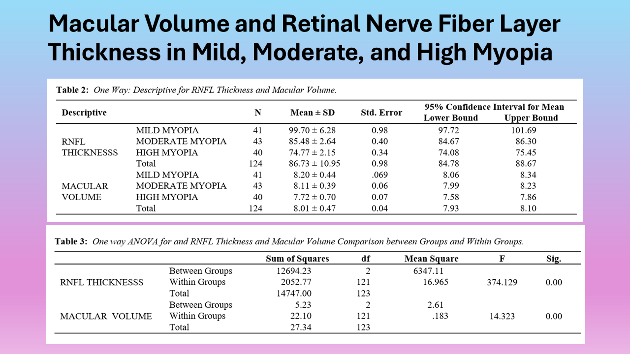
Methods: One hundred and twenty-four eyes were included in the study. Based on the spherical equivalent (SE) refractive error, participants were divided into three groups: moderate myopia (from -3.00 D to -6.00D), high myopia (<-6.00 D), and mild myopia (>-3.00 D and ≤ -0.50 D).Spectral-domain optical coherence tomography was used to evaluate thickness of RNFL and macular volume. For the statistical analysis one-way ANOVA and post-hoc Tukey HSD tests were employed.
Results: There were 69 males and 55 females. Age ranged between 18 and 45 years with average age of 30.96 years.The RNFL thickness varied significantly among myopia groups, as confirmed by the F-statistic (374.129) and p-value of < 0.001. There was significant thinning of RNFL with increase in myopia. Macular volume significantly differed among the three myopia groups as confirmed by the significant finding (F = 14.323,
p < 0.001). Post-hoc analysis showed that there was significant difference between macular volume of mild and high myopia(p < 0.001). As the severity of myopia increased, macular volume gradually decreased.
Conclusion: Decrease in macular volume was a gradual structural alteration. Significant RNFL thinning was observed in cases of severe myopia.
Downloads
Published
How to Cite
Issue
Section
License
Copyright (c) 2026 Athar Habib, Anila Farid, Muhammad Asim Saeedi, Shehar Yar Abdullah Sabir, Maher Mustansar Ali Qasim

This work is licensed under a Creative Commons Attribution-NonCommercial 4.0 International License.


