
Comparison of Corneal and Anterior Chamber Parameters between Myopic Eyes and the LASIK – Treated Eyes Using Pentacam
Doi: 10.36351/pjo.v42i1.2172
DOI:
https://doi.org/10.36351/pjo.v42i1.2172Abstract
Purpose: To compare the Corneal and Anterior Chamber Parameters between Myopic Eyes and the LASIK-treated eyes Using Pentacam
Study Design: Cross sectional observational study
Place and duration of study: Department of Optometry, College of Applied Medical Sciences, Qassim University from January 2024 to May 2024.
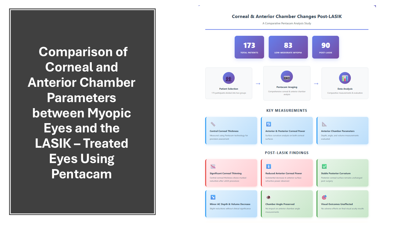
Methods: The study included 173 patients, 83 individuals with low to moderate myopia and 90 post-LASIK patients. Corneal and anterior chamber measurements were taken using Pentacam, including central corneal thickness, anterior and posterior corneal surface power, anterior chamber depth, angle, and volume.
Results: The mean spherical equivalent was -2.01 ± 1.02 D in myopic eyes and -0.51 ± 0.41 D in post-LASIK eyes (P = 0.000). Central and thinnest corneal thicknesses were significantly reduced in the post-LASIK group (P = 0.000). Uncorrected visual acuity in post-LASIK eyes (mean 1.03 ± 0.15) was comparable to the best-corrected visual acuity in myopic eyes (P = 0.098). A significant decrease in anterior corneal surface power was noted post-LASIK, while posterior corneal power remained stable. Anterior chamber depth and volume showed modest but significant reductions, with no significant difference in anterior chamber angle between the groups.
Conclusion: LASIK leads to significant thinning of the cornea and reduction in anterior corneal power, while posterior corneal curvature remains stable. Minor decreases in anterior chamber depth and volume occur without affecting the chamber angle or visual outcomes.
Downloads
Published
How to Cite
Issue
Section
License
Copyright (c) 2025 Majid A. Moafa

This work is licensed under a Creative Commons Attribution-NonCommercial 4.0 International License.


